

Up until now, making a piece of bone-like material to repair bone tissue of a patient involves first going into a laboratory to fabricate the structures using high-temperature furnaces and toxic chemicals.
A novel 3D printing method which can be utilized inside the patient’s body provides hope for better trauma and cancer treatment; in a nutshell, diseases and operations that require bone replacements, reducing pain and speeding up recovery time.
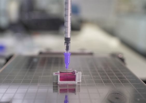
Called ceramic omnidirectional bioprinting in cell-suspensions (COBICS), the method is a result of a research carried out by scientists from UNSW Sydney who have developed a ceramic-based ink that may allow surgeons in the future to 3D-print bone parts complete with living cells that could be used to repair damaged bone tissue.
In other words, blending a ceramic material that mimics bone structure with the patient’s own cells in a 3D printing “ink”, may lead to the creation of new bone material inside the body. The latter can replace removed sections of bone and existing bones can be knitted with the new artificial bone.
This is not the first attempt of 3D-printing bone-mimicking structures. However, according to Dr Iman Roohani from UNSW’s School of Chemistry, the trial performed using a Lulzbot 3D printer reveals that for the first time, such material can be created at room temperature – complete with living cells – and without harsh chemicals or radiation.
Living cells can now be part of the 3D-printed structure
Until now, 3D printing of bones has involved producing material in a laboratory, exposing the structures to high-temperature furnaces and toxic chemicals.
“This produces a dry material that is then brought into a clinical setting or in a laboratory, where they wash it profusely and then add living cells to it,” explains Associate Professor Kristopher Kilian who co-developed the new process with Roohani.
“The cool thing about our technique is you can just extrude it directly into a place where there are cells, like a cavity in a patient’s bone. We can go directly into the bone where there are cells, blood vessels and fat, and print a bone-like structure that already contains living cells, right in that area”, he continues.
“The ink takes advantage of a setting mechanism through the local nanocrystallisation of its components in aqueous environments, converting the inorganic ink to mechanically interlocked bone apatite nanocrystals,” Dr Roohani says.
“In other words, it forms a structure that is chemically similar to bone-building blocks. The ink is formulated in such a way that the conversion is quick, non-toxic in a biological environment and it only initiates when ink is exposed to the body fluids, providing an ample working time for the end-user, for example, surgeons.”
While the new process has raised interest from surgeons and medical technology manufacturers who see big potential in a wide range of medical fields including dental restoration, the next step for the duo of scientists is to perform in vivo tests in animal models. The goal of this process is to discover if the living cells in the bone-like constructs continue to grow after being implanted in existing bone tissue.
Remember, you can post job opportunities in the AM Industry on 3D ADEPT Media free of charge or look for a job via our job board. Make sure to follow us on our social networks and subscribe to our weekly newsletter : Facebook, Twitter, LinkedIn & Instagram ! If you want to be featured in the next issue of our digital magazine or if you hear a story that needs to be heard, make sure you send it to contact@3dadept.com


