

Feature first published in the May/June 2022 issue of 3D ADEPT Mag and posted online in September 2022. Interviewee roles, forecasts and commercial examples reflect that period. Research demonstrations should not be confused with transplant-ready organs.
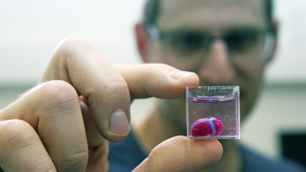
Whenever one reads an announcement that highlights 3D printed organs, one can easily be tempted to think that these organs can already be implanted within a human body but reality shows that there is a large gap between the capabilities and limitations encountered with 3D bioprinting.
3D bioprinting uses controlled deposition or patterning of biological materials, often including cells, to produce three-dimensional constructs. Printing a shape does not itself make a mature, functional tissue: culture, maturation and validation remain essential.
The field includes tissue models for research, drug testing and longer-term regenerative medicine goals. Some approaches may help reduce reliance on animal experiments, while food-related work follows different requirements. These aims should be distinguished from established clinical treatments.
“One of the ultimate goals of 3D bioprinting is to develop a viable solution that will be able to realize 3D functional complex organs but there is more than that. Research around 3D bioprinting has evolved and has started to move to industrial-based applications. One of the clear goals of 3D bioprinting is organ and tissue replacement. The other one is to create complex cell discoveries for making better medicine and quicker”, Dr. Simon Mackenzie, CEO of REGENHU told 3D ADEPT Media.
Yet, the reality, as per the words of Stephen Gray, cofounder of Ourobionics, is that “we should arrive to a solution where we merge human tissue with bioelectronics and biosensors to improve or to try to create models for drug discovery. Ultimately, we should be able to develop human electronics that could be used for implants or other medical devices. Realistically, printing an organ is a topic that we could [objectively] discuss in 10+ years, but exploring human tissue options with bioelectronics and sensors is something we can do now; it’s something that can lead to tangible improvements across segments such as drug discovery.”
“This means, the first area of focus right now is outside the body and the second stage will be replacing [or transferring] the applications inside the body” John Zandbergen, CEO of Ourobionics adds.
The “10+ years” estimate and development priorities are the interviewees’ views in 2022, not a promised clinical timetable.
By 2022, research had demonstrated a range of printed structures and tissue models, while major obstacles remained in function, scale and reproducibility.
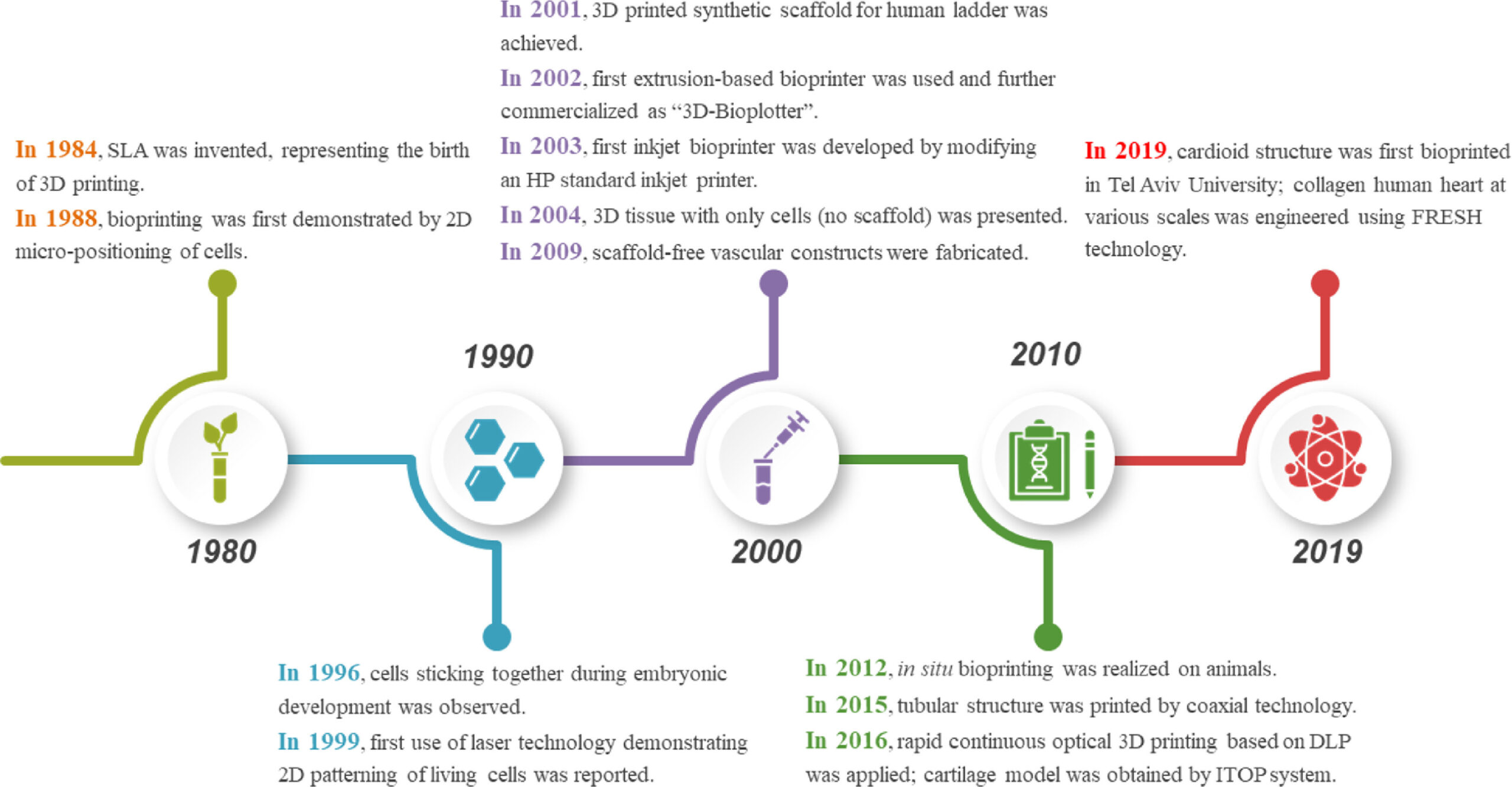
This feature examines the methods, limitations and priorities discussed by researchers and suppliers in 2022. The following historical timeline provides context for that discussion.
How does 3D bioprinting work?
Bioprinting draws on tissue engineering, cell biology, biomaterials, microfabrication and additive manufacturing. Equipment development and biological control must progress together.
The truth is, “the 3D printing world solved a lot of issues; we have learned [and we continue to learn] a lot from these companies. They may think it’s not their path, but they are wrong. We are just many years behind them”, Mackenzie outlines.
One of the first lessons the 3D printing world inspired, is a manufacturing process that follows the steps you probably already know: preparation (3D imaging, 3D modeling, Bioink preparation), printing process, post-bioprinting (cross-linking, maturation).
A process may start with medical imaging, a CAD model or another validated design. Planning software then defines deposition or exposure instructions for the selected system; not every bioprinter uses the same file format or layer strategy. After printing, cross-linking and controlled culture conditions can help stabilise and mature the construct. A bioreactor can reproduce selected environmental conditions, rather than the full complexity of an organism.
Needless to say, variations in these stages may occur depending on the type of 3D bioprinting process the operator leverages and as seen with conventional additive manufacturing processes, there are also several types of 3D bioprinting processes.
Four commonly described families are microextrusion, inkjet, laser-assisted deposition and stereolithography-based bioprinting. This is a useful framework, not an exhaustive or universally fixed classification.
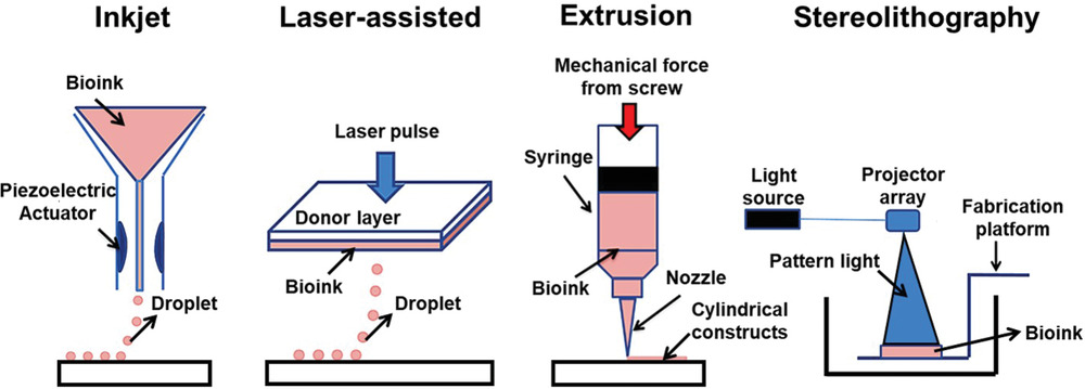
Each 3D bioprinting process comes with its share of advantages and limitations:
| Method | Principle | Potential advantages | Limitations |
|---|---|---|---|
| Microextrusion | Deposits bioink through a nozzle using pneumatic or mechanical force. | Can handle relatively viscous formulations and continuous structures. | Cell stress, nozzle blockage and achievable detail depend on ink, nozzle and settings. |
| Inkjet | Places droplets, commonly by thermal or piezoelectric actuation. | Rapid patterning and multiple dispensing heads. | Requires compatible viscosity, cell concentration and droplet stability. |
| Laser-assisted | Laser energy transfers material from a donor layer to a receiving surface. | No dispensing nozzle; fine placement can be possible. | Transfer conditions, ink properties and energy exposure can affect cell survival. |
| Stereolithography-based | Light patterns cross-link a suitable photosensitive formulation. | Fine geometry without nozzle extrusion. | Photochemistry, light dose, material compatibility and maturation constrain performance. |
This editorial comparison draws on the 2021 review, “3D Bioprinting at the Frontier of Regenerative Medicine, Pharmaceutical, and Food Industries”. It avoids universal cell counts per droplet, viscosity limits or resolution promises: these need a specified system and test method.
Extrusion and inkjet are widely used approaches, alongside hybrid and emerging methods. Electrohydrodynamic jetting uses an electric field to control deposition; melt electrowriting typically makes polymer scaffolds that can later be combined with cells. Magnetic assembly, acoustic methods, microneedle supports and embedded printing in support baths address different fabrication tasks. They should not all be treated as identical ways of directly printing living cells.
“Those other processes that are not often found in the main categories are much more niche technologies. They are often combined with extrusion and inkjet to create better processes”, the CEO of REGENHU explains.
At the end of the day, most of these processes (if not all of them) seem to be trying to solve a pivotal issue during the printing process:
“One of the main problems with 3D bioprinting is that everything gets damage during the process. In order to create human tissue, you need something that keeps the cells alive otherwise they don’t form the tissue you are looking for. With extrusion bioprinting you can get some basic structures, but you can’t go to the next level”, Stephen Gray, cofounder of Ourobionics told 3D ADEPT Media.
This is Gray’s assessment in the 2022 interview. Cell survival depends on the cell type, material and operating conditions; damage is not inevitable in every process.
Another aspect we should take into account is the level of complexity, Zandbergen says. “You cannot create different kinds of complexity with the same tissue. Each tissue presents its array of limitations depending on its purpose. The level of complexity you are able to create inside the 3D structure determines the level of functionality of the tissue you are making. Don’t expect a huge level of complexity when it comes to the depth of the tissue when using basic bioprinting processes”, he points out.
So, what are some of the key criteria that may lead to the choice of one process over another?
The choice should begin with the biological question and the function to be reproduced. The interviews highlight cell compatibility, geometry, time and final purpose as interdependent criteria.
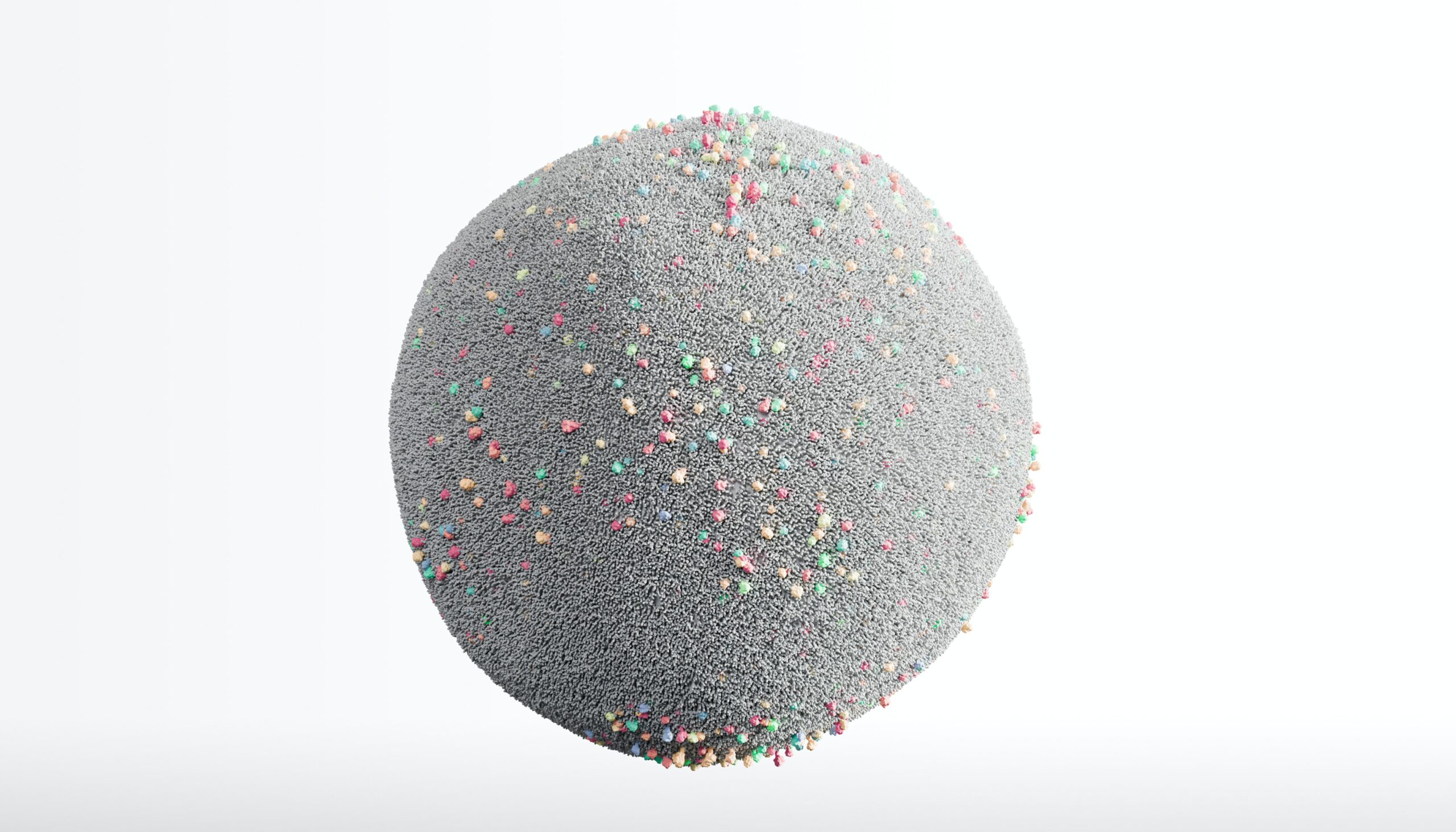
The first one is cells. As speakers from Ourobionics said, the goal here is to keep the cells alive while working on them. Tissue printing requires a large number of cells. “It’s a very crucial problem because you cannot create a human tissue if everything is dying in the process. One of the recent improvements in this area has been to add a volumetric principle to the printing process of some technologies. Light-based technologies are also getting to a level where they are slightly better but the problem here remains inside the resin – which in my opinion – cannot deliver a functional organ”, Zandbergen lays emphasis on.
Speed is another criterion, but faster deposition is not always better. Printing time, exposure and mechanical stress must be balanced against cell survival and construct stability. Maturation can then change cell distribution, tissue organisation and mechanical behaviour.
The final purpose determines the appropriate compromise. A printer’s positioning specification does not establish the resolution of a cell-laden construct. No general 1 µm precision can be promised for syringe extrusion. Cell viability, geometry, reproducibility and function should be measured for the intended research or clinical application.
Applications and areas for improvement
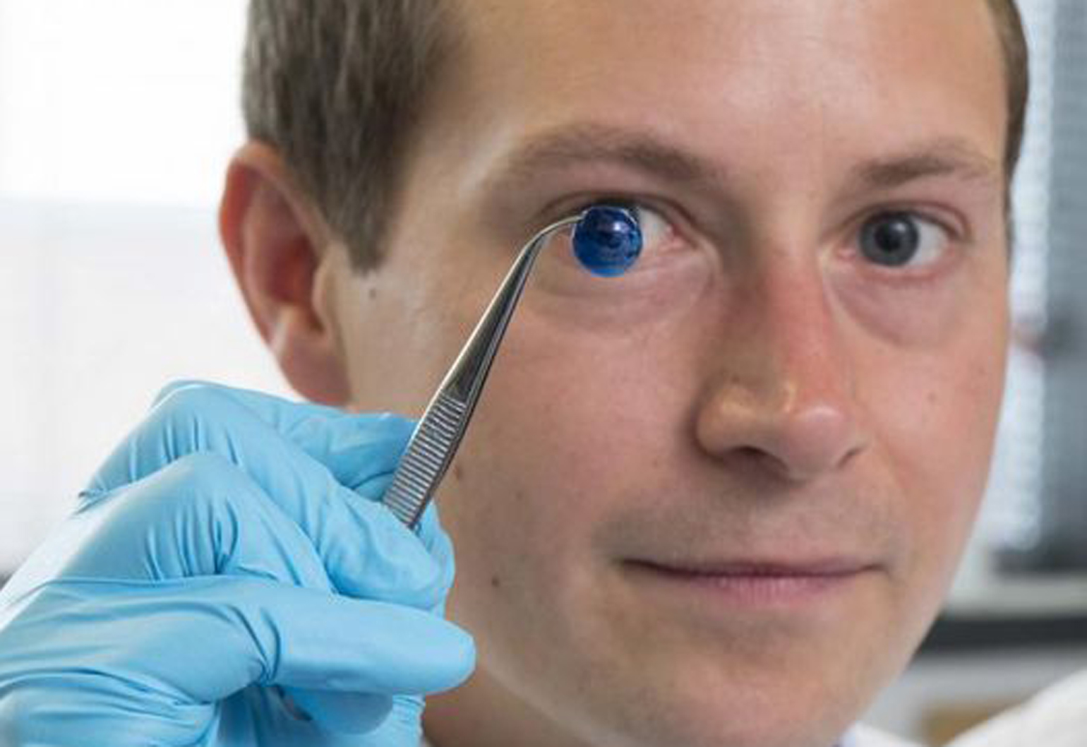
The Newcastle study published in 2018 demonstrated a corneal stroma equivalent. Its feasibility result should not be read as a cornea already available for patient transplantation.
“While many technologies touch upon organ and tissue replacement, we are still very far away from commercial applications. On the current market, the cell biology side is gaining momentum, especially for complex cell models as they ambition to reduce the need to use animals for testing products. Applications that are closed to market include the printing of beta cells for diabetes”, Mackenzie’s points out.
These comments describe the speaker’s outlook in 2022. “Close to market” does not identify a regulatory authorisation or establish routine patient access.
Bioprinted models can help investigate cell interactions, disease mechanisms and responses to candidate drugs. How closely a model reproduces a human organ must be demonstrated for its intended experiment; shape and the presence of cells alone do not establish physiological equivalence.
In situ bioprinting is another research direction, including approaches to deposit skin-related cells and materials at a wound site. The example covered by 3D ADEPT concerns research, rather than a generally available treatment.
Food applications require a distinction between cultivated meat made from animal cells and plant-based products shaped by 3D printing. MeaTech was pursuing cell-based meat development, whereas Redefine Meat’s European restaurant launch in 2021 concerned plant-based products. That launch is not evidence that bioprinted cultivated meat was commercially available in those restaurants.
However, there are a number of limitations that remain to address to help the market move forward efficiently:
| Area | Issue to address |
|---|---|
| Process complexity | Integrate equipment, cells, materials and reproducible protocols; setup and validation can take substantial time. |
| Biological suitability | Design printable materials that support the intended cell behaviour and tissue environment. |
| Material and equipment compatibility | Match rheology, cross-linking and mechanical properties to the selected printing system. |
| Software and control | Plan complex structures and monitor the relevant fabrication and biological parameters. |
| Data interoperability | Improve the exchange of geometry, process settings and supporting biological information. |
| Cost and access | Assess equipment, consumables, training and validation costs against the research objective. Extrusion remains useful where it meets that objective. |
Editorial synthesis of the development priorities discussed with REGENHU and Ourobionics in 2022.
Concluding thoughts
Bioprinting deserves coverage both for demonstrated research applications and for the obstacles that remain. A useful assessment separates laboratory results, industrial tools, clinical investigations and authorised treatments.
The shortage of donor organs is a major motivation for this work. Waiting-list figures vary by country and date; the original article’s undated numerical comparison is not retained as a current measure.
That unmet need does not make a printed organ an available alternative. Progress requires convincing evidence of tissue function, vascularisation, reproducibility and safety, followed by the appropriate clinical and regulatory pathway. These are the priorities against which future announcements should be assessed.
This dossier has first been published in the May/June 2022 edition of 3D ADEPT Mag.
Resources and contributors
REGENHU, founded in 2007, develops bioprinting equipment. The R-GEN 100 and R-GEN 200 systems were among the company’s offerings discussed for this 2022 feature.
Ourobionics describes a development approach combining biofabrication with bioelectronics, biosensors and therapeutic agents. Its stated ambition in the interview was to support regenerative medicine, medical devices and human–machine interfaces; these aims do not establish clinical availability.
3D Bioprinting at the Frontier of Regenerative Medicine, Pharmaceutical, and Food Industries, Front. Med. Technol., 28 January 2021 | https://doi.org/10.3389/fmedt.2020.607648
Development of 3D bioprinting: From printing methods to biomedical applications, research, Asian Journal of Pharmaceutical Sciences
3D Bioprinting of Lignocellulosic Biomaterials, https://doi.org/10.1002/adhm.202001472
THE 3D ADEPT WEEKLY NEWSLETTER
The industry moves fast. Stay ahead.
Every week, the latest additive manufacturing news, in-depth features and real-world applications, straight to your inbox. Plus, be the first to know when a new issue of 3D ADEPT Mag is out.
Free · One email a week · Unsubscribe anytime
Clicking loads our email provider’s sign-up form, including its analytics and Google’s anti-bot protection. You’re only subscribed if you complete the sign-up. Read our privacy policy.