Among the various birth defects that might require an in-utero surgery, there is spina bifida, a condition that can happen anywhere along the spine if the neural tube does not close all the way. Such condition can result in a lifetime of neurological disabilities, including the inability to walk.
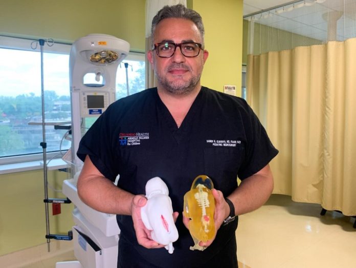
To help address this issue, surgeons from Orlando Health Winnie Palmer Hospital for Women & Babies combine MRI and ultrasound imaging with 3D printing to create a detailed model that enabled them to prepare for a procedure that could treat this condition. In this specific case, the models aim to help identify hurdles and reduce risks of surgery.
“The 3D reconstruction of the fetus can really educate the surgeon on the real-life shape, size and location of the spinal lesion, as well as prepare the surgeon to have the appropriate equipment ready to treat this condition surgically. It’s a level of detail that we are not able to see in traditional imaging, but that is extremely valuable in these cases where we cannot actually see the defect ahead of surgery“, Samer Elbabaa, MD, Medical Director, Pediatric Neurosurgery, Orlando Health explains.
From a technological standpoint, the medical team worked with the 3D printing experts from Digital Anatomy Simulations for Healthcare, LLC (DASH). The company’s technology has improved MRI and ultrasound images taken throughout the pregnancy and makes it easy to reconstruct accurate curves and edges.
Thereafter, they 3D printed the high-resolution model with multiple colours and materials. The result enables doctors to clearly assess items such as skeletal structure, nerve and vascular anatomy and fluid sacs in the spine and brain caused by spina bifida.
“The fetal models not only help surgeons plan for things like where to make an incision and how to repair the defect, but also helps reduce the duration of the surgery to limit the developing baby’s exposure,” said DASH President and CEO Jack Stubbs.
“We are able to create models that are extremely realistic by using a stack of two-dimensional images taken throughout the pregnancy and enhancing them to reconstruct a better visualization of what the fetus truly looks like.”
“At first, we just thought it was a model showing the same kind of condition that our baby was diagnosed with, but then Dr. Elbabaa told us that it was made using the 20-week MRI of our daughter,” Jared Rodriguez, one of the parents said. “We could see the brain and the spine and I looked down at it and thought, ‘I’m holding my daughter right now? That’s pretty awesome.'”
The parents had envisioned the challenges their daughter may face and they can’t be more excited today for the healthier future she will have thanks to such technological advancements.
According to the medical experts, most babies who undertake this procedure before they are born, have less health issues than those who experience surgery after their birth.
Source: News Medical – Remember, you can post free of charge job opportunities in the AM Industry on 3D ADEPT Media or look for a job via our job board. Make sure to follow us on our social networks and subscribe to our weekly newsletter : Facebook, Twitter, LinkedIn & Instagram ! If you want to be featured in the next issue of our digital magazine or if you hear a story that needs to be heard, make sure to send it to contact@3dadept.com


