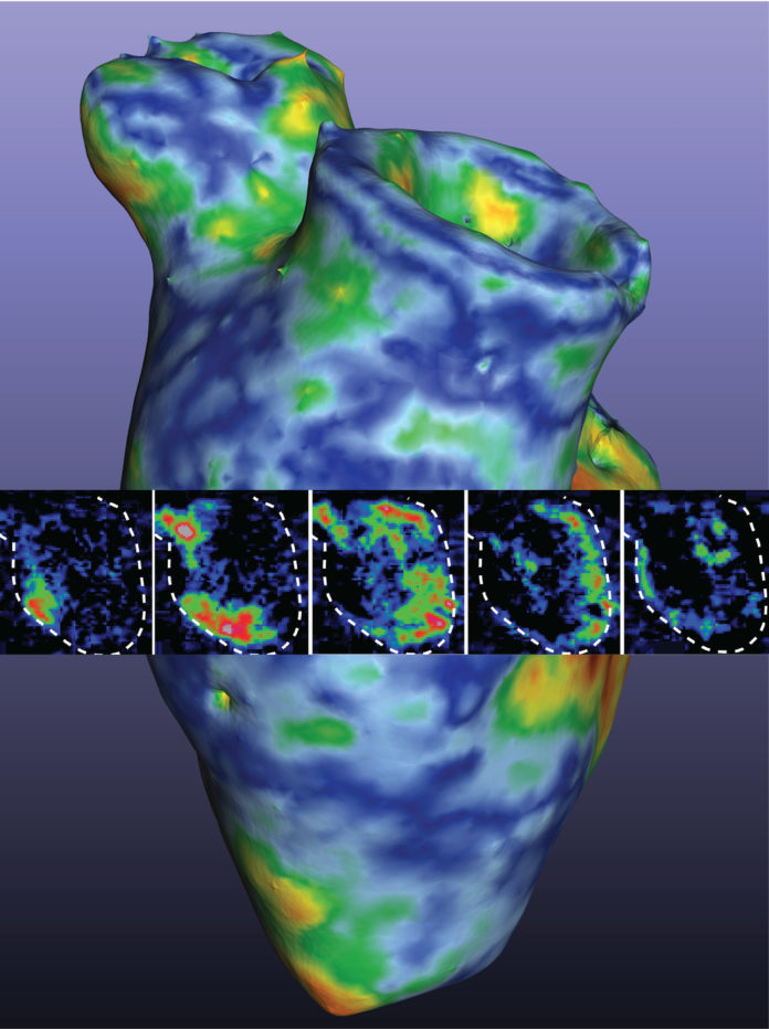
Researchers from the University of Minnesota have 3D printed a functioning centimeter-scale human heart pump, an innovation that will improve research for heart diseases.
In the past, scientists have tried to 3D print heart muscle cells (called cardiomyocytes) that were derived from what are called pluripotent human stem cells. These are cells that have the potential to develop into any type of cell in the body. The idea was to reprogram these stem cells to heart muscle cells before 3D printing them to obtain a three-dimensional structure, called an extracellular matrix. The problem with this method was that it was not possible to reach critical cell density for the heart muscle cells to actually function.
After flipping the process…
“At first, we tried 3D printing cardiomyocytes, and we failed, too,” said Brenda Ogle, the lead researcher on the study and head of the Department of Biomedical Engineering in the University of Minnesota College of Science and Engineering. “So, with our team’s expertise in stem cell research and 3D printing, we decided to try a new approach. We optimized the specialized ink made from extracellular matrix proteins, combined the ink with human stem cells and used the ink-plus-cells to 3D print the chambered structure. The stem cells were expanded to high cell densities in the structure first, and then we differentiated them to the heart muscle cells.”
For the first time ever, they could achieve the goal of high cell density within less than a month to allow the cells to beat together, just like a human heart.
“After years of research, we were ready to give up and then two of my biomedical engineering Ph.D. students, Molly Kupfer and Wei-Han Lin, suggested we try printing the stem cells first,” said Ogle, who also serves as director of the University of Minnesota’s Stem Cell Institute. “We decided to give it one last try. I couldn’t believe it when we looked at the dish in the lab and saw the whole thing contracting spontaneously and synchronously and able to move fluid.”

According to the lead scientist, these heart muscle cells work in a way that the cells could organize and work together. With about 1.5 centimeters long, the heart muscle model can fit into the abdominal cavity of a mouse for further study.
“We now have a model to track and trace what is happening at the cell and molecular level in pump structure that begins to approximate the human heart,” Ogle said. “We can introduce disease and damage into the model and then study the effects of medicines and other therapeutics.”
The full study can be read on Science Daily.
Remember, you can post AM job opportunities for free on 3D ADEPT Media or look for a job via our job board. Make sure to follow us on our social networks and subscribe to our weekly newsletter: Facebook, Twitter, LinkedIn & Instagram! If you want to be featured in the next issue of our digital magazine or if you hear a story that needs to be heard, make sure to send it to contact@3dadept.com


