From one research to another, 3D printing might enable to open new fields or create new 3D printing techniques. Remember how researchers from Illinois developed free-form printing to 3D print sugar scaffolds. Anyway, this week also starts with a new 3D printing technique developed by MIT and Harvard in the field of science.
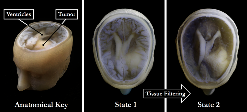
This time, science was inspired by the personal story of Ph.D student Steven Keating whose tumor was removed from his brain when he was 26. This phase of his life made him curious to discover what his brain looked like before the tumor was removed, the goal being to better understand the diagnosis and possibilities of treatment.
So, how did he do that?
After collecting his medical data, the researcher began 3D printing his MRI and CT scans after realizing that, the first results with existing methods were not that conclusive. Another method which consists in 3D printing biological samples demonstrated significant results.
This research resulted in an impromptu collaboration which gathered together James Weaver, Ph.D., Senior Research Scientist at the Wyss Institute; Neri Oxman, Ph.D., Director of the MIT Media Lab’s Mediated Matter group and Associate Professor of Media Arts and Sciences; and a team of researchers and physicians at several other academic and medical centers in the US and Germany.
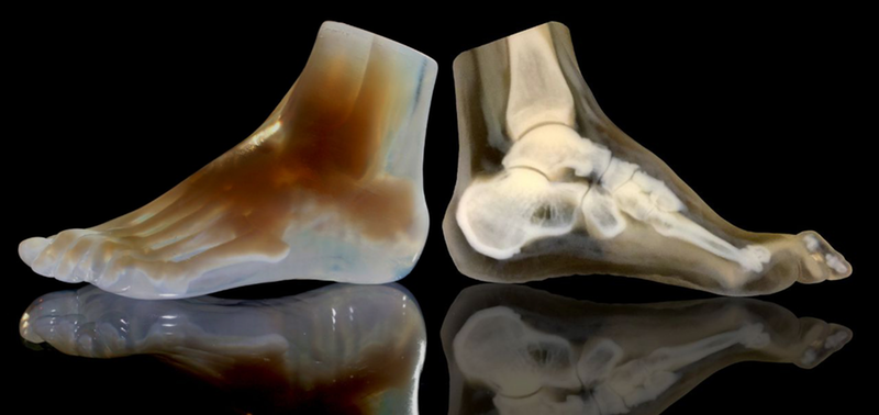
The technique they have developed enables “images from MRI, CT, and other medical scans to be easily and quickly converted into physical models with unprecedented detail.”
Imaging technologies like MRI and CT scans produce high-resolution images as a series of “slices” that reveal the details of structures inside the human body, making them an invaluable resource for evaluating and diagnosing medical conditions. Most 3D printers build physical models in a layer-by-layer process, so feeding them layers of medical images to create a solid structure is an obvious synergy between the two technologies.
The only challenge was that MRI and CT scans produce very detailed images, therefore, the part that must be 3D printed needs “to be isolated from surrounding tissue and converted into surface meshes in order to be 3D printed.”

Researchers achieved it via the “segmentation” which consists in tracing the desired object on every single image slice. They could also proceed by the automatic “thresholding” process. In this case, areas with grayscale pixels are turned into either solid black or solid white pixels, based on a shade of gray that is chosen to be the threshold between black and white.
 “I nearly jumped out of my chair when I saw what this technology is able to do,” says Beth Ripley, M.D. Ph.D., an Assistant Professor of Radiology at the University of Washington and clinical radiologist at the Seattle VA, and co-author of the paper. “It creates exquisitely detailed 3D-printed medical models with a fraction of the manual labor currently required, making 3D printing more accessible to the medical field as a tool for research and diagnosis.”
Imaging technologies like MRI and CT scans produce high-resolution images as a series of “slices” that reveal the details of structures inside the human body, making them an invaluable resource for
This new method gives medical experts the best of both worlds: a fast and highly accurate method for converting complex images into a format that can be easily 3D printed.
It also shows us how far curiosity can bring us.
For further information about 3D Printing, follow us on our social networks and subscribe to our newsletter!
Would you like to be featured in the next issue of our digital magazine? Send us an email at contact@3dadept.com
//pagead2.googlesyndication.com/pagead/js/adsbygoogle.js
(adsbygoogle = window.adsbygoogle || []).push({});


