Applications of 3D printing in the medical industry include both medical cares provided to human and to animals. A recent interesting example of treatment integrating 3D printing offered to an animal is the integration of an experimental biomimetic prosthesis into a dog with osteosarcoma of the femur.
For those who do not know osteosarcoma also known as osteogenic sarcoma is a fast-growing cancer whose cells originate from bone tissue, that results in a gradual destruction and sometimes to the loss of mobility. Both humans and animals might suffer from this disease.
The experimental biomimetic prosthesis
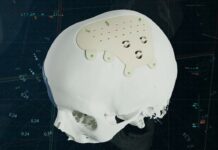
A prosthesis that is biomimetic is similar in structure to the tissues of a living organism. To treat this cancer, chemotherapy and surgery are necessary to remove the affected tissues. Even if doctors get accustomed to insert a metal, ceramic or polymer implant into the bone, it should be noted that, while restoring mobility, prostheses are very different in structure from bone tissue, therefore may create a number of issues.

“The traditional materials for medical prosthetics have a number of significant disadvantages: for example, titanium implants take on too much of the load intended for the bone, and the latter begins to thin out. In this situation, the bone at the junction with the prosthesis may break. Another option is ceramics, but they are more fragile, which may limit the size of the recovered bone tissue. In addition, the structure of these materials does not allow them to ‘grow together’ with the bone – a constant tight fixation is required,” explained Fedor Senatov, General Director ofBiomimetix and research assistant at the NUST MISIS Center of Composite Materials.
“Since the dog was big, it used to move [very much], so 11 cm of bone was required to be removed, and the prosthesis [needed to be] a hybrid. On the titanium tube, made with 3D printing by ‘Konmet’, our partners, we have increased the layer of solid ultrahigh molecular weight polyethylene, and the inner part is made of porous ultrahigh molecular weight polyethylene, identical to the structure of the spongy bone. During the operation, part of the coating was cut to ‘fit’ the implant to the bone. A few days after the surgery, the dog was able to walk. If the fusion of the polymer andbone tissue is successful, it will be possible to remove the fixing plates after some time,” – said Senatov.
Further information has not been given regarding 3D printing. However, we do know that the implant is durable, as it corresponds to the weight of the dog and the bone size. For now, the dog is undergoing chemotherapy. Doctors are satisfIed with the results of the operation and expect a lot of progress.
For further information about 3D Printing, follow us on our social networks and subscribe to our newsletter
Would you like to subscribe to 3D Adept Mag? Would you like to be featured in the next issue of our digital magazine? Send us an email at contact@3dadept.com


